- 현재 위치
- home > 인체모형/실습모형/CPR > 심장모형/폐,간,신장모형 > [3B] 동맥,정맥확대모형 (G42)
; "확대")


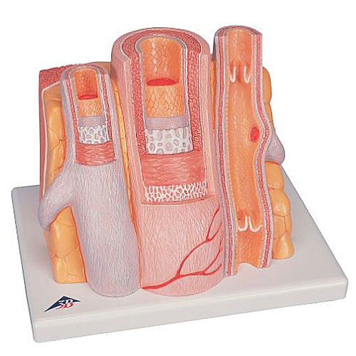
동맥, 정맥 14배 확대 모형
이 모형은 14배로 확대된 근육과 조직에 인접한 전완 근처의 정맥이 있는 중간 사이즈의 근육 동맥을 보여준다.
The MICROanatomy™ circulatory system model illustrates the reciprocal anatomical relationship of artery and vein and the basic functional techniques of the venous valves (“valve function” and “muscle pump”).
The left vein and the middle artery are fenestrated in the upper anterior segment, revealing the various layers of the wall structure in a cross and longitudinal section and in top view.
The right vein is opened throughout in the anterior segment, revealing the orifice of a feeder vein and two venous valves, i.e. “flap valves” formed by a duplication of the tunica intima.
On the rear of the artery and vein model, the relief of two veins is shown to illustrate the functional aspect of the venous valves.
A great tool for teaching about the human circulatory system. Artery and vein supplied on base.
26 x19 x18,5 cm : 0,9 kg 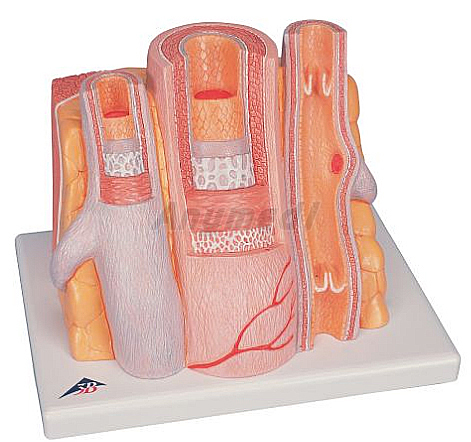


 |